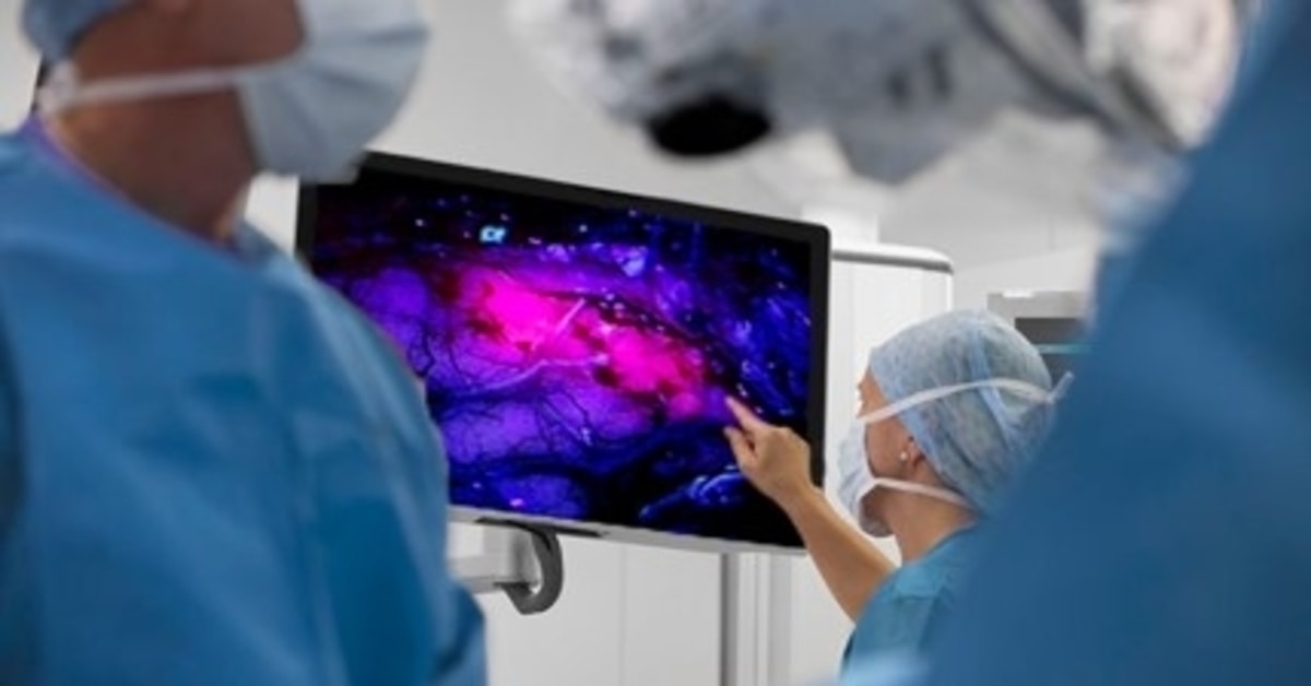
The neurosurgeons at BayCare Clinic have a new way to see brain tumors.
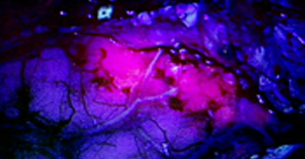
Gleolan is a dye that illuminates tumors during brain surgery. The tumors turn bright pink or magenta under a special blue light. The color distinction helps neurosurgeons better identify the tumor from healthy brain matter, ensuring a more thorough tumor resection or removal and better outcome for the patient.
Patients ingest the dye 2 to 4 hours before surgery.
The dye is specifically used to help neurosurgeons see brain tumors called high-grade gliomas. These life-threatening tumors can spread into brain tissue, making them difficult to remove or resect.
Before Gleolan, this form of brain surgery would be performed under white light, making it challenging for neurosurgeons to differentiate between diseased and healthy tissue.
“The Gleolan dye is used to light up a brain tumor while we work to remove it,” says Dr. Max Ots, a neurosurgeon with BayCare Clinic Neurological Surgeons. He was the first BayCare Clinic surgeon to successfully use the Gleolan dye during brain surgery. “It helps to more accurately define the margins of the tumor to help ensure more aggressive resection.”
Gleolan does that by binding to a protein found only in glioma tumors. The cancerous areas glow when viewed under a specially-filtered microscope, Ots says. The dye fades after a few hours.
“Having this new imaging tool should give our brain surgery patients added comfort when heading into a surgical procedure,” Ots says. “We have the ability to better map the delineation of malignant tissue during brain surgery. That should lead to better outcomes for our patients.”
After surgery, patients using Gleolan must remain in a dark environment for up to three days.